Vestibular diagnostics at a glance · Gaze stabilisation
DVA
Dynamic visual acuity · visual acuity during head movement
The vHIT measures the vestibulo-ocular reflex – the DVA shows what it achieves for vision. Patients with a VOR deficit see blurred during head movements or complain of oscillopsia. The DVA makes this measurable: as the loss of visual acuity between a still and a moving head, in logMAR.
Landolt ring per ISO 8596Passive head impulses above 150 °/sResult in logMARAdds vision to the vHIT
How the test works All tests Technical information (PDF, German)
What the test measures
Gaze stabilisation – as visual acuity
First the static visual acuity (SVA) is determined with the head still. Then the optotype appears only while the head is moved quickly – the dynamic visual acuity (DVA).
The difference is the DVA loss in logMAR: 0.1 logMAR corresponds to one line of acuity, 0.2 logMAR roughly to a drop from 1.0 to 0.63 decimal acuity.
In head impulse DVA a Landolt ring appears only when a passive head impulse exceeds a velocity threshold. The ring size adapts. Visual acuity drops measurably from about 2–3 °/s of retinal slip.
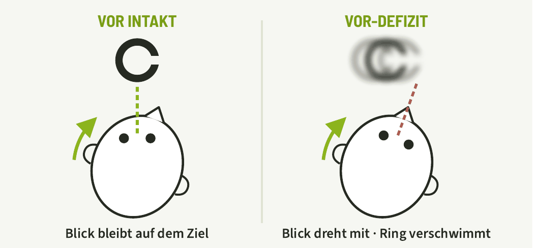
Schematic, head seen from above, green arrow = head impulse. Graphic from the DVA technical information.
How the test works
Patient seated · own spectacle correction · video goggles with head motion sensor
1
Prepare
The patient sits in front of the screen at the defined distance, with usual correction. Explain how to respond: state the direction of the ring opening.
2
Static visual acuity
Landolt rings with the head still, adaptively smaller down to threshold. Gives the reference value in logMAR.
3
Recognition time
Determine the shortest display time the patient reliably recognises, so that slow processing is not rated as a VOR deficit.
4
Head impulses
The examiner delivers small, fast, unpredictable passive impulses. The ring appears only above the threshold (e.g. 150 °/s).
5
Evaluate
Determine DVA per impulse direction. DVA loss = DVA − SVA; right/left comparison.
Passive instead of active: with active, predictable head movements the brain can counteract with pre-programmed eye movements – a deficit then appears smaller. Passive high-velocity impulses separate healthy subjects and patients much better (Vital 2010; Herdman 2003).
Evidence
High diagnostic accuracy
100 % / 94 %
Sensitivity / specificity of head impulse DVA vs. search-coil HIT (n = 115)
94,5 % / 95,2 %
Sensitivity / specificity of computerised DVA (n = 97)
- Good reproducibility (ICC 0.83–0.87)
- Separates healthy subjects, unilateral and bilateral loss (p < 0.001)
- DVA loss increases slightly with age
Clinical benefit
When the DVA helps
- Objectify oscillopsia – the key symptom of bilateral vestibulopathy
- Bárány criteria: DVA loss ≥ 0.2 logMAR is considered pathological (supplementary test)
- Function behind the vHIT finding: shows whether gain deficit and saccades impair vision in everyday life
- Therapy monitoring: in randomised trials, vestibular training improved DVA in unilateral and bilateral loss
Classification
DVA in the test battery
| vHIT | DVA | SVA | |
|---|---|---|---|
| Question | Reflex gain | Vision during head movement | Vision at rest |
| Measure | Gain, saccades | logMAR loss | logMAR |
| Side | yes | yes | – |
| Cooperation | low | response needed | response needed |
Limitations: requires sufficient visual acuity, cooperation and cognition. Values depend on age and test setup. The DVA complements the vHIT but does not replace it.
Result
What you see in the result
- Static and dynamic visual acuity in logMAR
- DVA loss per impulse direction, right/left comparison
- Patient's recognition time
- Report together with the vHIT result
Complements
vHIT
Measures the reflex directly (gain, saccades) – the DVA shows the effect on vision.
SHIMP
Second head impulse paradigm, e.g. for follow-up.
VertiGuard
Balance during everyday movements – the other side of everyday function.
With Merlin
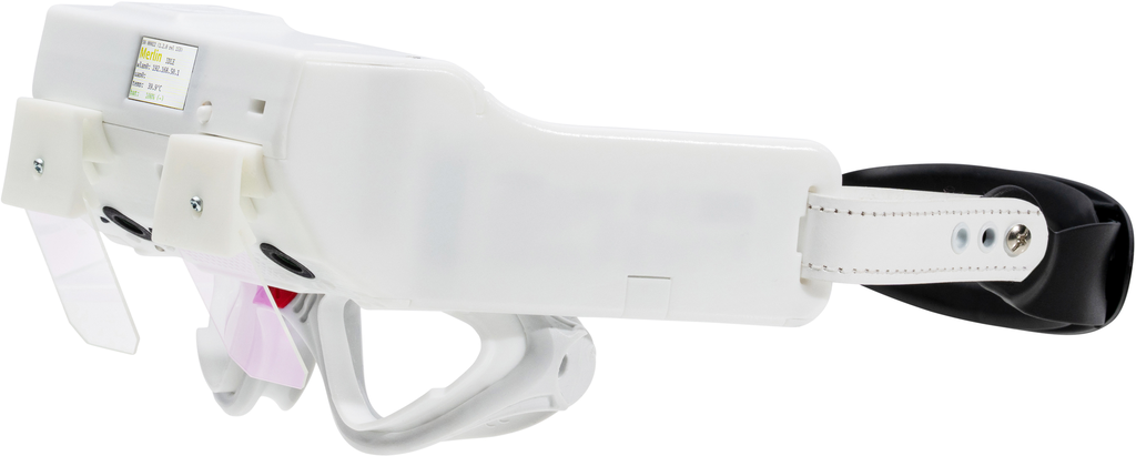
The DVA is a software module for Merlin. medicAide Nexus shows the Landolt rings on a screen, the goggles' motion sensor measures head velocity.
| Module | Merlin DVA |
|---|---|
| Included in | Merlin Clinical, Merlin Complete |
| Requires | Merlin video goggles + medicAide Nexus with screen |
| Individually on demand (pay-per-use) | €11.90 per examinationAvailable – price on request |
Prices incl. 19% VAT (Germany). On demand: one examination per patient per day; requires a maintenance contract or a subscription on the system.
Frequently asked questions
With active, predictable movements the brain can counteract with pre-programmed eye movements. Passive, unpredictable high-velocity impulses reveal a VOR deficit much more reliably.
According to the Bárány criteria for bilateral vestibulopathy, a DVA loss of 0.2 logMAR or more is considered pathological (supplementary test). Values depend on age and setup.
Sufficient visual acuity with own correction and the ability to state the direction of the ring opening.
No. The vHIT measures the reflex, the DVA its effect on vision. Together they give the finding and its everyday consequence.
References
- Vital D, Hegemann SC, Straumann D, et al. A new dynamic visual acuity test to assess peripheral vestibular function. Arch Otolaryngol Head Neck Surg 2010;136(7):686–691.
- Herdman SJ, Tusa RJ, Blatt P, et al. Computerized dynamic visual acuity test in the assessment of vestibular deficits. Am J Otol 1998;19(6):790–796.
- Herdman SJ, Schubert MC, Das VE, Tusa RJ. Recovery of dynamic visual acuity in unilateral vestibular hypofunction. Arch Otolaryngol Head Neck Surg 2003;129(8):819–824.
- Herdman SJ, Hall CD, Schubert MC, et al. Recovery of dynamic visual acuity in bilateral vestibular hypofunction. Arch Otolaryngol Head Neck Surg 2007;133(4):383–389.
- Demer JL, Honrubia V, Baloh RW. Dynamic visual acuity: a test for oscillopsia and vestibulo-ocular reflex function. Am J Otol 1994;15(3):340–347.
- Strupp M, Kim JS, Murofushi T, et al. Bilateral vestibulopathy: diagnostic criteria. Consensus document of the Bárány Society. J Vestib Res 2017;27(4):177–189.
DVA technical information (PDF, German)
Technical information for physicians. For billing questions, please contact your billing service.
See the DVA live?
We will show you the test in a free online demo – with your questions.