Vestibular diagnostics at a glance · Central or peripheral?
Oculomotor testing
Gaze holding · saccades · smooth pursuit · optokinetics
Vestibular tests examine the labyrinth, oculomotor testing examines control by the brainstem and cerebellum. Four short tests with the video goggles help to separate peripheral from central causes – measured instead of just observed.
Four testsBrainstem and cerebellumStimuli on screenComplements the vHIT
How the test works All tests Technical information (PDF, German)
The four tests
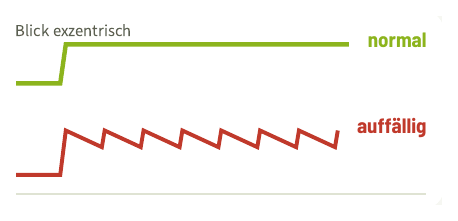
Gaze-holding test
Gaze holding · nystagmus
Tests
Are the eyes stable in eccentric gaze? Is there spontaneous nystagmus?
Abnormal
Gaze-evoked nystagmus: drift to the centre, corrective saccades. In all directions → cerebellum, medication, alcohol.
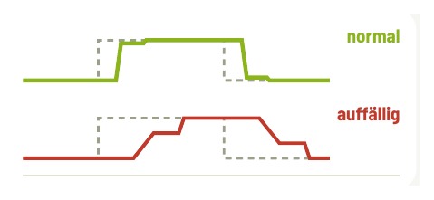
Saccades
Saccades
Tests
Latency, velocity and accuracy of fast gaze shifts.
Abnormal
Slowed or dysmetric with corrective saccade. Slow only vertically → midbrain.
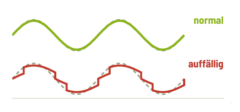
Smooth pursuit
Smooth pursuit
Tests
Does the eye follow a slow target smoothly? Gain = eye ÷ target.
Abnormal
Saccadic: the eye lags behind and catches up with saccades. Asymmetric → central sign.
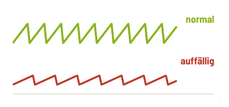
Optokinetics
Optokinetic nystagmus
Tests
Reflex nystagmus in response to a large moving stripe pattern.
Abnormal
Slow phase weak or asymmetric, often together with impaired smooth pursuit.
Stimulus / target Eye, normal Eye, abnormal · schematic eye position over time
How the test works
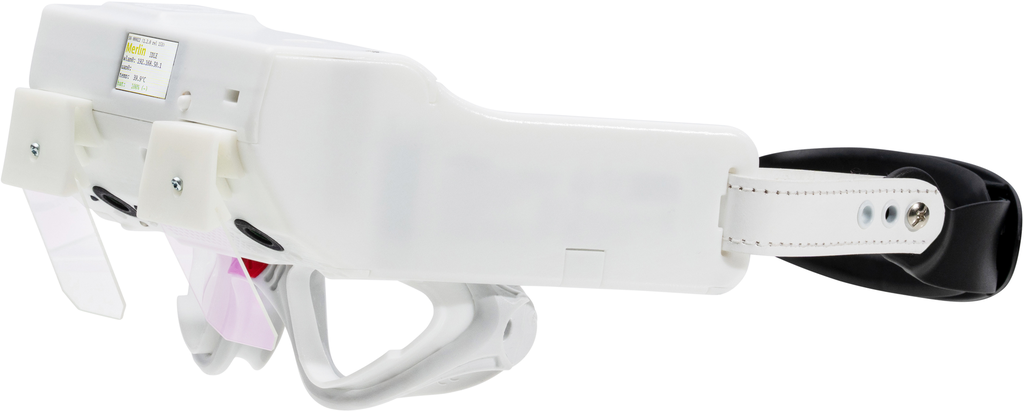
Patient seated · head still · stimuli on screen · video goggles
1
Set up
Fixed distance to the screen, head still. Calibrate eye position with fixation points – deflections in degrees.
2
Gaze holding
Fixate straight ahead, right, left, up, down – about 20–30° eccentric, not at end position – and hold.
3
Saccades
A dot jumps between positions, ideally with random amplitude and timing. Test horizontally and vertically.
4
Smooth pursuit
The dot moves slowly back and forth in a sine wave. The patient follows with the eyes only; gain and saccades per direction.
5
Optokinetics
Stripe pattern across the whole visual field, to the right and left. Evaluate slow phase and side comparison.
Interpret correctly: an acute peripheral loss can also disturb smooth pursuit and optokinetics – but this is compensated quickly. Persistent disturbances and saccadic abnormalities indicate a central cause. Therefore record spontaneous nystagmus first.
Evidence
Measured, not estimated
≈ 80 %
of PSP patients identified via vertical saccades and intrusions; specificity above 95% (n = 100)
34 %
of MS patients with INO, objectively via saccade measurement (n = 210)
- Smooth pursuit separates MSA (catch-up saccades) from Parkinson's disease (anticipatory saccades)
- Cerebellum: gaze-evoked nystagmus from 20°
Clinical benefit
When oculomotor testing helps
- Vertigo of unclear cause: detect central signs, in addition to the vHIT
- Chronic dizziness, gait unsteadiness: detect cerebellar signs
- Neurological diseases: signs in MS (INO), PSP, MSA, ataxias
- Follow-up: measurements instead of impressions, for reports and referrals
Topographic diagnosis
Finding and lesion site
| Finding | Points to |
|---|---|
| Gaze-evoked nystagmus in all directions | Cerebellum, medication |
| Only vertical / only horizontal gaze-evoked nystagmus | Midbrain / pontomedullary |
| Downbeat nystagmus | Flocculus bilaterally |
| Slowed vertical saccades | Midbrain (e.g. PSP) |
| Slowed horizontal saccades | Pons (PPRF) |
| Slowed adduction during saccades | MLF: INO (e.g. MS) |
Limitations: all parameters depend on age – use age-related norms. Alertness, cooperation and medication have an influence.
Result
What you see in the result
- Eye position over time per test, stimulus and eye overlaid
- Saccades: latency, velocity, accuracy
- Smooth pursuit: gain per direction, saccadic intrusion
- Optokinetics: slow phase and side comparison
- PDF report for findings and referral
Complements
vHIT and SHIMP
Test peripheral function – together with oculomotor testing they distinguish peripheral from central.
Positional and positioning tests
Detect positional nystagmus and distinguish it from central patterns.
VNG / caloric testing
Spontaneous nystagmus and side-specific excitability.
With Merlin
The oculomotor test battery is the OKM software module for Merlin. medicAide Nexus shows the stimuli on a screen – no extra PC needed.
| Module | Merlin OKM |
|---|---|
| Included in | Merlin Complete |
| Requires | Merlin video goggles + medicAide Nexus with screen |
| Individually on demand (pay-per-use) | €23.80 per examinationAvailable – price on request |
Prices incl. 19% VAT (Germany). On demand: one examination per patient per day; requires a maintenance contract or a subscription on the system.
Frequently asked questions
Gaze holding, saccades, smooth pursuit and optokinetics.
No. medicAide Nexus shows the stimuli on a screen; the patient sits in front of it at a fixed distance.
Yes, an acute peripheral loss can temporarily impair smooth pursuit and optokinetics. This is compensated quickly. Persistent disturbances and saccadic abnormalities indicate a central cause.
Yes, also on a Screening or Clinical system with medicAide Nexus – on demand for individual patients.
References
- Strupp M, Kremmyda O, Adamczyk C, et al. Central ocular motor disorders, including gaze palsy and nystagmus. J Neurol 2014;261 Suppl 2:S542–558.
- Wunderlich J, Behler A, Dreyhaupt J, et al. Diagnostic value of video-oculography in progressive supranuclear palsy: a controlled study in 100 patients. J Neurol 2021;268(9):3467–3475.
- Nij Bijvank JA, van Rijn LJ, Balk LJ, et al. Diagnosing and quantifying a common deficit in multiple sclerosis: internuclear ophthalmoplegia. Neurology 2019;92(20):e2299–e2308.
- Pinkhardt EH, Kassubek J, Süssmuth S, et al. Comparison of smooth pursuit eye movement deficits in multiple system atrophy and Parkinson’s disease. J Neurol 2009;256(9):1438–1446.
- Tarnutzer AA, Weber KP, Schuknecht B, et al. Gaze holding deficits discriminate early from late onset cerebellar degeneration. J Neurol 2015;262(8):1837–1849.
- Baloh RW, Honrubia V, Sills A. Eye-tracking and optokinetic nystagmus: results of quantitative testing in patients with well-defined nervous system lesions. Ann Otol Rhinol Laryngol 1977;86:108–114.
- Seferlis F, Chimona TS, Papadakis CE, et al. Age related changes in ocular motor testing in healthy subjects. J Vestib Res 2015;25(2):57–66.
- Leigh RJ, Zee DS. The Neurology of Eye Movements. 5. Aufl. Oxford University Press 2015.
Oculomotor technical information (PDF, German)
Technical information for physicians. For billing questions, please contact your billing service.
Oculomotor testing in your practice?
We will show you the test in a free online demo – with your questions.